Glaucoma vs Cataract: Why an Eye Pressure Test Isn’t Enough
I want to start with the sentence that gave me a false sense of security for about three years.
“Your pressure looks fine.”
That was the report my mother brought home from every eye appointment, and I took it to mean the same thing she did — that glaucoma had been checked for and ruled out. It hadn’t been. A pressure reading is one number from one test, and that test misses roughly half of the people who have the disease.
So this is the article I wish someone had handed us. Glaucoma and cataract are two completely different problems — one is a clouding you can see through less and less, the other is a quiet narrowing at the edges that you don’t notice until a lot of it is gone. They get lumped together because both arrive with age and both are found in the same chair, in the same clinic, on the same afternoon.
Here’s what each one actually is, why a normal pressure number proves almost nothing, what a real exam includes, how often to actually go, and the one version of this that is a genuine emergency.
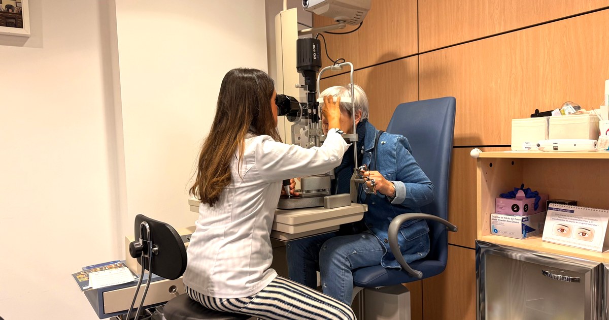
Photo: José Antonio Otegui Auzmendi / Pexels
Two different parts of the eye, two different problems
The simplest way to hold these apart is to remember which piece of hardware is failing.
Cataract is a clouding of the lens. The National Eye Institute describes the mechanism plainly: around age 40, the proteins in the lens start to break down and clump together, and the clouding worsens over time. Cataracts can also follow an eye injury, eye surgery, or radiation. It is a light-transmission problem. The window fogs up.
Glaucoma is damage to the optic nerve. The nerve fibers carrying the picture from your eye to your brain die off. Open-angle glaucoma is the most common form in the U.S.; angle-closure and congenital glaucoma are less common. This is a wiring problem, and wiring does not grow back.
That single distinction predicts almost everything else about them.
| Cataract | Glaucoma | |
|---|---|---|
| What’s damaged | The lens clouds over | The optic nerve fibers die |
| Early symptoms | Often none; later blurry vision, faded colors, glare, halos, poor night vision | Usually none at all until loss is extensive |
| Reversible? | Yes — surgery removes the clouded lens; 9 of 10 people see better | No — existing damage is permanent; treatment stops it worsening |
| The test that finds it | Dilated eye exam | Dilated optic nerve exam + visual field + OCT |
| Urgency | Not urgent — waiting typically doesn’t cause harm | Time-sensitive. Acute angle-closure: emergency, hours matter |
Cataract is extremely common, and that’s worth saying without alarm. More than 24.4 million Americans age 40 or older have cataracts — about 17% of that age group. The rate climbs steeply: roughly 2.5% at 40–49, about 15.5% in the early 60s, 36.5% at 70–74, 49.5% at 75–79, and more than half of Americans by age 80.
Glaucoma is less common but far more silent. About 4.22 million people in the U.S. have it, and 1.49 million have vision-affecting glaucoma. Among adults 40 and older, 2.56% have glaucoma. And per CDC figures, about half of those affected don’t know it — one analysis put the number of people with undetected, untreated glaucoma at roughly 2.4 million.
Half. That’s the whole reason this article exists.

Cloudy vision is one thing. Vision quietly narrowing is another.
Cataract announces itself, eventually. Early on there are often no symptoms at all, but later come blurry or cloudy vision, faded colors, trouble seeing at night, light sensitivity, halos around lights, and double vision. It is annoying in a way you can describe to another person.
Glaucoma does not do that. Early open-angle glaucoma typically has no warning signs whatsoever. The AAO puts it bluntly: many people are unaware they have it until it causes significant, irreversible vision loss, because the disease often has no symptoms until the loss is already extensive.
Now, a nuance that matters, because the standard picture of glaucoma is misleading.
You’ve probably seen the illustration — a black tunnel closing in from the edges. The general pattern is real enough: peripheral vision is commonly affected first, often starting near the nose, and central vision may hold up until later stages. But researchers have flagged the tunnel image as a commonly held misconception. Except in very advanced disease, patients describe their field loss as blurred areas, not black holes. The symptoms people actually report most often are needing more light and blurry vision.

Photo: SHVETS production / Pexels
There’s more. Recent anatomical and psychophysical evidence supports meaningful central visual dysfunction earlier than the conventional teaching assumed, and standard perimetry optimized for peripheral loss may overestimate peripheral loss while underestimating central loss.
So the honest version is this: the early damage is usually off to the side, and you experience it as dim or blurry patches you never consciously register — because your other eye and your brain fill in the gap.
That is exactly why it goes undetected. Nothing hurts. Nothing looks obviously wrong. And half of the people who have it never find out until an exam finds it.
One overlap worth knowing: halos
Both conditions can produce halos around lights, which confuses people. The difference is the company they keep.
Cataract halos creep in gradually alongside glare and night-driving trouble. Halos that arrive suddenly with severe eye pain, nausea, and a red eye are a different animal entirely — that’s the emergency at the end of this article.
Why a normal pressure reading doesn’t rule out glaucoma
This is the part that changed how I read my mother’s appointment notes.
The belief that glaucoma equals high eye pressure is not just imprecise. It’s wrong often enough to be dangerous.
Normal-tension glaucoma is glaucomatous optic nerve damage with pressure in the normal range — conventionally an IOP of 21 mmHg or below, a threshold chosen because about 95% of normal subjects fall between 11 and 21 mmHg. The European Glaucoma Society’s 2021 guidelines define it as a specific type of open-angle glaucoma with nerve head damage and matching visual field defects in patients whose pressure stays consistently under 21.
How big a share is that? Averaged across patients diagnosed with a glaucomatous visual field defect, normal-tension glaucoma accounts for 30–40% of cases, and epidemiologic estimates suggest 50% or more of all open-angle glaucoma worldwide. In people of White or European ancestry it’s about a third of open-angle glaucoma; in sub-Saharan African ancestry, around half.
And then there’s the finding that I think should be printed on the wall of every waiting room. In the Baltimore Eye Survey, half of participants with primary open-angle glaucoma had an IOP below 21 mmHg at the time of diagnosis — and 20% of that subset were under 21 at each of their first three visits.
Not one low reading. Three.
NEI states the underlying reason simply: normal eye pressure varies by person, and what’s normal for one person could be high for another. On top of that, the measurement itself is shaky. Thin corneas cause applanation tonometry to underestimate pressure, corneal hysteresis adds more error, and pressure fluctuates through the day — which is why specialists sometimes measure across 24 hours rather than trusting a single office reading.
The numbers that settle it
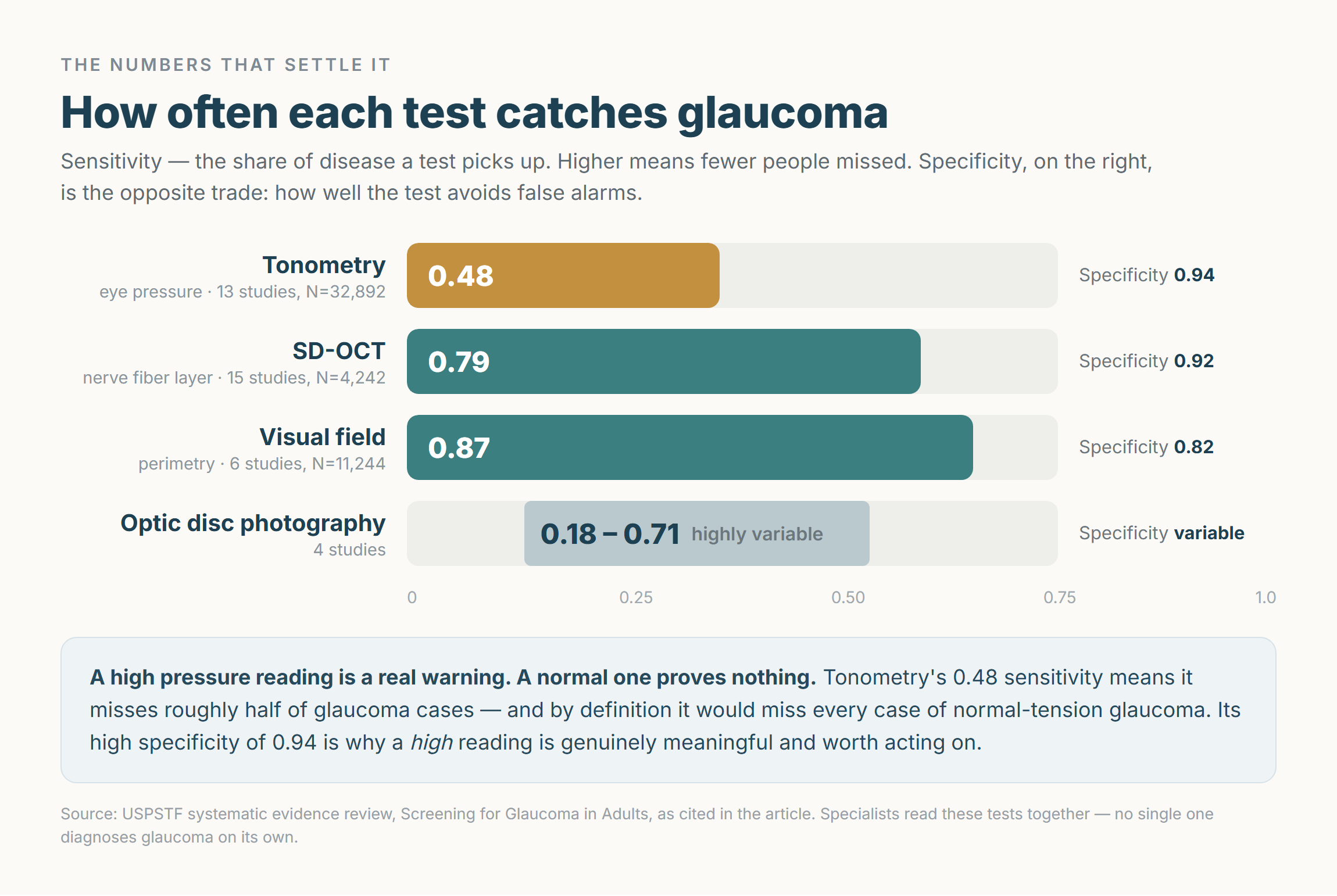
The USPSTF’s systematic evidence review pooled the diagnostic accuracy of each test. This table is the practical heart of the whole topic.
| Test | Sensitivity (catches disease) | Specificity | Evidence base |
|---|---|---|---|
| Tonometry (eye pressure) | 0.48 | 0.94 | 13 studies, N=32,892 |
| Visual field | 0.87 | 0.82 | 6 studies, N=11,244 |
| SD-OCT (nerve fiber layer) | 0.79 | 0.92 | 15 studies, N=4,242 |
| Optic disc photography | 0.18–0.71 (highly variable) | variable | 4 studies |
Read the top row twice. A sensitivity of 0.48 means tonometry misses roughly half of glaucoma cases. Its high specificity of 0.94 means the opposite is also true — a high reading is genuinely meaningful and worth acting on.
So the takeaway isn’t “pressure tests are useless.” It’s asymmetric:
A high pressure reading is a real warning. A normal one proves nothing.
And by definition, a pressure test alone would miss every single case of normal-tension glaucoma.
What a real glaucoma exam actually includes
The standard workup is not one test. It’s a set, and specialists read them together before diagnosing anything.
- Tonometry — measures eye pressure. Finds a major treatable risk factor, not the disease itself.
- Dilated optic nerve exam — the clinician gets a magnified, 3D view of the optic nerve. This is where the actual damage, the cupping and rim thinning, is seen. It requires dilation, which is why the drops matter.
- Visual field test (perimetry) — maps your complete field of vision and reveals blind spots you cannot perceive on your own. Highest sensitivity of the group at 0.87, but it detects functional loss that has already happened.
- OCT — cross-sectional imaging of the retinal nerve fiber layer. Detects structural thinning often before the visual field changes, at 0.79 sensitivity. This is the test that most changed early detection.
- Gonioscopy — determines whether the drainage angle is open and wide or narrow and closed. In other words, it identifies who is at risk of the emergency form.
- Pachymetry — measures corneal thickness. The landmark Ocular Hypertension Treatment Study confirmed corneal thickness is an independent risk factor for developing glaucoma, and it also corrects the pressure reading.
Which gives you the one sentence to bring to your next appointment.
Don’t ask for a pressure check. Ask for a comprehensive dilated eye exam that includes an evaluation of the optic nerve — and if you have risk factors, expect visual field testing and OCT as well. A pressure reading at a kiosk, or as a quick add-on during a general physical, is not a glaucoma screen.

Photo: VICTOR REGA / Pexels
So how often should you actually go?
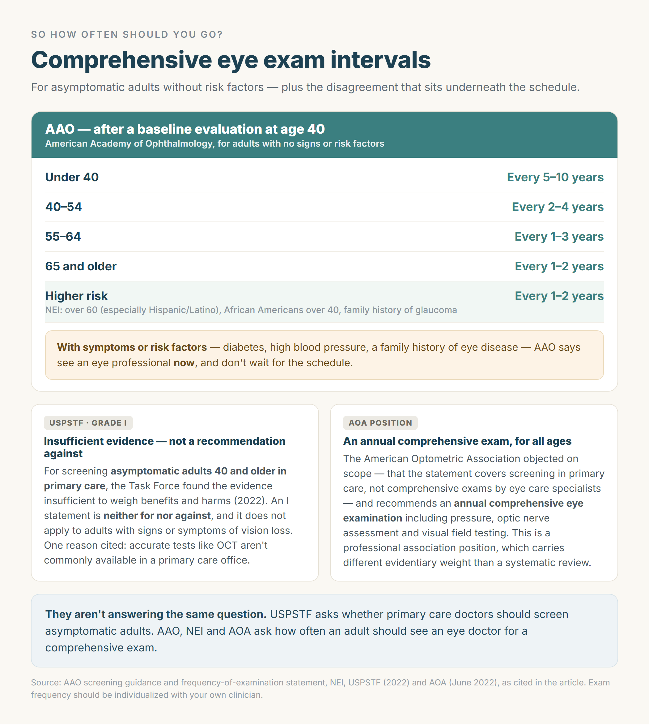
Here’s where two legitimate organizations say different-sounding things, and it’s worth understanding why before you decide anything.
AAO: a baseline at 40, then intervals by age and risk
The American Academy of Ophthalmology recommends that adults with no signs or risk factors get a comprehensive medical eye evaluation at age 40 if they haven’t had one, because early signs of disease and changes in vision often begin around then.
After that baseline, for asymptomatic adults without risk factors:
| Age | Interval |
|---|---|
| Under 40 | Every 5–10 years |
| 40–54 | Every 2–4 years |
| 55–64 | Every 1–3 years |
| 65+ | Every 1–2 years |
Which means the widely repeated line — “everyone over 40 needs an annual pressure and fundus exam” — overstates it. That is not what AAO recommends. (These interval figures come from AAO’s public guidance on eye disease screening and its clinical statement on frequency of ocular examination, corroborated across two AAO-sourced pages.)
The exceptions matter more than the schedule, though. AAO says people with symptoms or risk factors — diabetes, high blood pressure, a family history of eye disease — should see an eye professional now and should not wait.
NEI agrees on the higher-risk track: if you’re at higher risk, get a comprehensive dilated eye exam every 1 to 2 years. NEI defines higher risk as people over 60 (especially Hispanic/Latino individuals), African Americans over 40, and anyone with a family history of glaucoma. For cataract specifically, NEI advises adults 60 and older get an exam every 1–2 years.
USPSTF: insufficient evidence — and what that actually means
The U.S. Preventive Services Task Force gives glaucoma screening a grade I (insufficient evidence), finalized in 2022 and consistent with its earlier 2013 statement. Its conclusion: the current evidence is insufficient to assess the balance of benefits and harms of screening for primary open-angle glaucoma in adults.
Read the scope carefully, because it’s the whole story. The statement covers asymptomatic adults 40 and older, and it explicitly does not apply to adults with reported signs or symptoms of vision loss, or with other diagnosed types of glaucoma. An I statement is neither for nor against — clinicians use judgment for individual patients.
The reasons USPSTF gave are revealing:
- No direct evidence that screening improves actual health outcomes like vision function and quality of life, even though treatment improves intermediate markers.
- Limited evidence that improving those intermediate markers translates into better health outcomes.
- Accurate tests like OCT are not commonly administered in the primary care setting.
- Insufficient evidence on risk-assessment tools for identifying who benefits.
- Higher prevalence in Black and Hispanic/Latino populations was acknowledged, but adequate evidence on screening those higher-risk groups wasn’t found.
- Potential for overdiagnosis and overtreatment.
The eye care professions pushed back. In June 2022, the American Optometric Association raised a scope objection — that the recommendation applies only to screening in primary care settings, not to comprehensive eye exams performed by eye care specialists, and that screenings should be used solely to refer patients for a comprehensive exam, never to replace one. One optometrist quoted in that response argued the earlier 2013 I statement may itself have discouraged the very screening that would have generated the missing evidence, a kind of catch-22. Another warned that primary care physicians may read an I statement as “glaucoma is low priority,” reducing referrals.
The AOA’s own position is more aggressive than AAO’s: an annual comprehensive eye examination for all age populations, including history, pressure measurement, optic nerve assessment, and visual field testing.
I’m not going to pick a winner here, because they aren’t answering the same question.
USPSTF asks: should primary care doctors screen asymptomatic adults? Answer: we don’t have the trial evidence to say.
AAO, NEI, and AOA ask: how often should an adult see an eye doctor for a comprehensive exam? Answer: baseline at 40 and a schedule by age and risk, or annually.
Two honest observations to hold alongside that. An I statement means “we don’t know,” not “don’t get your eyes checked” — and notably, one of the strongest reasons USPSTF cited is that OCT and optic nerve evaluation aren’t available in a primary care office, which is an argument for seeing an eye care professional rather than against it. At the same time, AOA’s annual recommendation is a professional association position, which carries a different kind of evidentiary weight than a systematic review does.
What cataract surgery realistically does
Since cataract is the fixable one, let’s be precise about what “fixable” means.
Timing is decided by function, not by how the cataract looks. NEI’s framing is that surgery becomes appropriate when the cataract interferes with daily activities like reading, driving, or watching television — and that “waiting typically won’t cause harm.” NHS-affiliated patient information says the same: surgery is usually only recommended if the loss of vision is affecting daily activities.
On effectiveness, I want to quote NEI exactly rather than paraphrase upward: “9 out of 10 people who get it can see better afterwards.”
See better. Not “restored to normal.” That distinction is the whole point.
The main limit is other eye disease. NHS-affiliated guidance notes that if you have another condition affecting your eyes, such as diabetes or glaucoma, you may still have limited vision even after successful surgery. Peer-reviewed data agrees — patients with coexisting eye disease have worse outcomes because of lower potential for visual function, and macular degeneration specifically relates to inferior outcomes for visual acuity and satisfaction compared with patients who have no coexisting eye disease.
But — and this is the part that gets left out — those patients still benefit. AMD patients gained in visual acuity and vision-related quality of life, and post-hoc AREDS analyses found most improved six months after surgery regardless of AMD severity.
The clouded lens is replaced with an artificial intraocular lens (IOL). Which type is a decision for you and your surgeon based on your individual eye, and I’m deliberately not going to compare them here. NEI describes the surgery as “very safe” without listing specific figures, so I won’t supply any either.
The two can coexist — and surgery nudges pressure down
Both are age-related, so plenty of people have both. There’s an encouraging wrinkle here.
Cataract surgery lowers eye pressure. Studies in glaucoma patients show a reduction of 2–4 mmHg lasting at least three years. Patients with primary open-angle glaucoma, or with narrow angles and chronic angle closure, were more likely to achieve a sustained reduction, and those with higher baseline pressure had increasingly higher odds of a reduction. In severe and end-stage primary angle-closure glaucoma with controlled pressure, cataract surgery significantly improved final visual acuity, and the number of eyes able to come off medications increased by 57.8%. MIGS combined with cataract surgery reduced pressure more than cataract surgery alone and cut postoperative medication use.
The honest caveat: the specific ocular characteristics that predict who gets this pressure-lowering benefit remain unclear, so it can’t be promised to any individual.
Treat it as a favorable side effect your surgeon factors into timing — not as a reason to seek cataract surgery to treat glaucoma. That’s a clinician’s judgment call.

Photo: cottonbro studio / Pexels
Irreversible, but not hopeless
Optic nerve damage doesn’t come back. NEI says treatment won’t undo damage to your vision, but it can stop it from getting worse. NHS-affiliated guidance says the same.
That sounds bleak until you look at what treatment does.
In the Early Manifest Glaucoma Trial, 255 open-angle glaucoma patients were randomized to treatment or no immediate treatment. Treatment halved the risk of progression (hazard ratio 0.50) — progression occurred in 45% of the treated group versus 62% of controls. Progression risk fell by about 10% for each 1 mmHg of pressure reduction from baseline, and when progression did occur in treated patients, it was delayed by a median of 18 months.
Now the counterweight, because “halved” sounds bigger than the reality. The USPSTF meta-analysis of topical medical therapy found a relative risk of 0.68 across 7 trials and 3,771 participants — an absolute risk reduction of about 4.8%, with one trial finding only non-significant differences in overall visual field loss. Relative risk reduction always sounds larger than the absolute benefit. Both numbers are true.
On blindness — two datasets, both real
The research on this genuinely conflicts, and I’d rather show you the disagreement than pick the comforting number.
On the reassuring side: in a retrospective study of 3,359 patients under clinical care with both eyes followed, 5.2% were predicted to progress to statutory blindness in their lifetime, with a further 10.4% reaching visual impairment. Outcomes have also improved — the likelihood of losing sight in one eye was 28% for patients diagnosed between 1965 and 1980, falling to 13.5% for those diagnosed between 1981 and 2000.
On the more pessimistic side: a Swedish cohort found that at the last visit, 42.2% had at least one blind eye from glaucoma and 16.4% were bilaterally blind — roughly one in six going bilaterally blind. Another analysis put cumulative incidence of glaucoma blindness in at least one eye at 26.5% at 10 years and 38.1% at 20 years after diagnosis.
The gap between 5.2% and 16.4% is largely explained by cohort era, how much damage existed at diagnosis, and length of follow-up. The older cohorts that drive the higher figures were diagnosed and treated in an earlier era of management.
So the fair synthesis: untreated, glaucoma ends in blindness. Caught early and treated consistently, most people keep useful vision for life — but “most” is not “all,” and the earlier it’s found, the better the odds.
The part that quietly decides the outcome
Drops only work if they go in the eye.
Reported nonadherence in glaucoma therapy ranges enormously, from 5% to 80%, depending on how it’s measured. About one third of people prescribed eye drops for the first time stop collecting prescriptions within a year. A University of Michigan study found 56% of glaucoma patients were non-adherent, with median monthly adherence of 74%.
And the reasons are mostly mechanical, not moral. Forgetfulness was the most common at 43.8%. Among the 62% who reported problems following the prescribed therapy, the leading obstacles were difficulty administering the drop (44%), cost (41%), and side effects (16%).
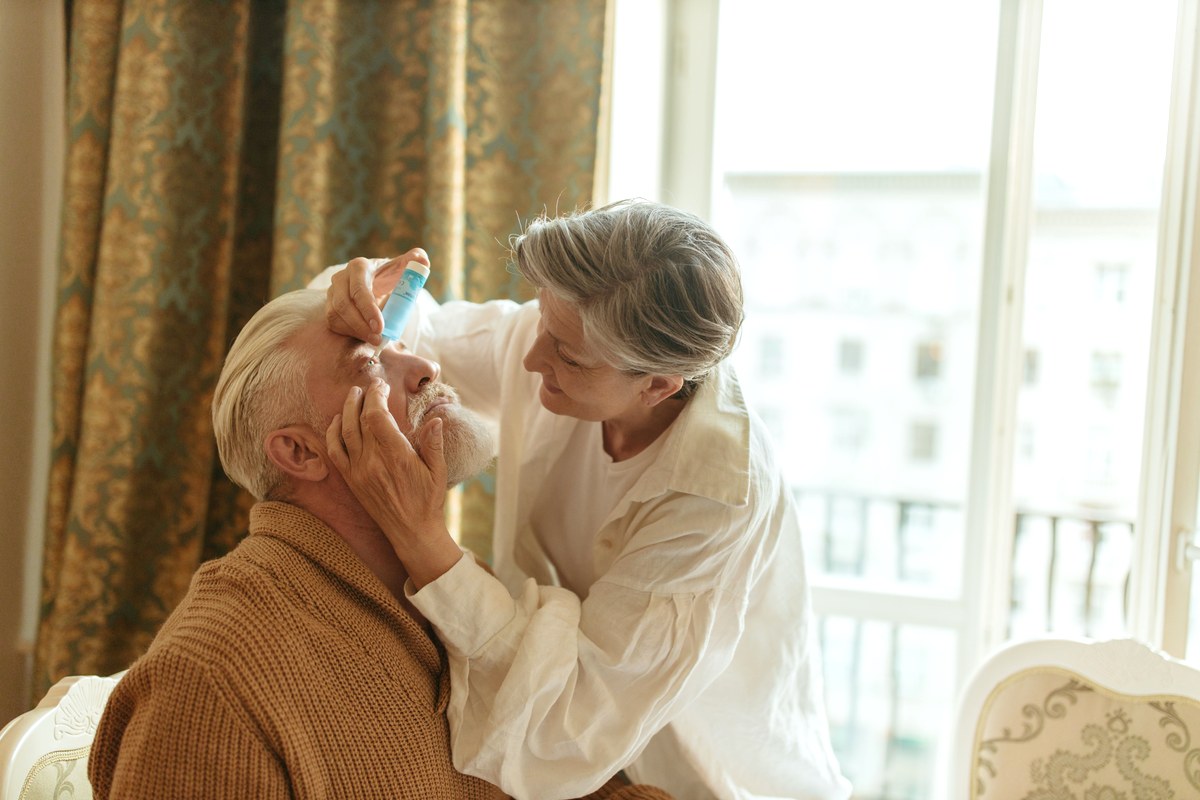
Photo: Yaroslav Shuraev / Pexels
If you cannot reliably get a drop into your own eye, that is a physical problem worth raising with your doctor — technique, drop aids, formulation, other options. It is not a character flaw, and it is not something to quietly give up on. Any change to a prescribed medication is a conversation with the prescribing clinician, not a decision to make alone.
The one eye emergency
Everything above is about time measured in years. This part is measured in hours.
Acute angle-closure glaucoma comes on suddenly with:
- Severe eye pain
- Headache
- Nausea and vomiting
- Halos or rainbow-colored rings around lights
- A red eye
- Sudden blurred vision
During an attack, eye pressure can climb to two or three times the normal level.
Why the clock matters: when pressure is lowered within the first few hours, many patients retain good visual function, but delays of even 12 to 24 hours can lead to significant, permanent vision loss. It is a medical emergency because permanent damage can happen very quickly, and it can lead to irreversible blindness without immediate treatment.
There’s a misdiagnosis trap here that I think is the single most useful warning in this article. Attacks can present with headache and abdominal pain with vomiting, and have been mistaken for gastroenteritis. If someone has a sudden severe headache and vomiting plus a red, painful eye and halos around lights, that is an eye emergency, not a stomach bug.
Risk rises with age, and is higher in women and in people of Asian descent. Angle-closure glaucoma affects roughly 1 in 1,000 people, with an estimated 17.14 million cases worldwide among people over 40.
What the ER or a glaucoma specialist does about it — pressure-lowering medication, positioning, laser peripheral iridotomy to let fluid flow freely — is entirely their job, and none of it is something to attempt or request by name.
Your instruction is one line long: go to an emergency room or contact a glaucoma specialist immediately. Any sudden vision loss deserves the same urgency.
What I’d tell my mother now
Cloudy vision and shrinking side vision are not two versions of the same problem. One is a lens that fogged over and can be replaced, on your schedule, when it starts getting in the way of reading and driving. The other is a nerve quietly losing fibers, with no symptoms, in about half of cases at a pressure reading that looks perfectly fine.
“Your pressure looks fine” answers one question, and it isn’t the question we thought it was answering.
The ask is small. A comprehensive dilated eye exam, with someone actually looking at the optic nerve — a baseline at 40, then on the schedule that fits your age and your risk. If glaucoma runs in your family, if you have diabetes or high blood pressure, if you’re Black and over 40 or Hispanic/Latino and over 60, ask your eye doctor for a shorter interval than the general schedule.
Nothing about this is urgent in the way an emergency is urgent. That’s exactly the problem with it — nothing ever feels like the day to go.
Medical disclaimer. This article is general health information, not medical advice, and it is not a diagnostic tool. Diagnosing glaucoma or cataract requires an in-person examination by an eye care professional. Do not start, stop, change, or skip any eye medication based on anything you read here — discuss adherence and any difficulty using drops with your prescribing clinician. Exam frequency should be individualized. Seek emergency care immediately for sudden severe eye pain, headache with nausea or vomiting, halos around lights, a red eye, or sudden blurred or lost vision — hours matter.
References
- National Eye Institute (NIH) — Cataracts; Glaucoma
- U.S. Preventive Services Task Force — Primary Open-Angle Glaucoma: Screening (Grade I, 2022); Screening for Glaucoma in Adults: A Systematic Evidence Review
- American Academy of Ophthalmology — Get an Eye Disease Screening by Age 40; Frequency of Ocular Examination clinical statement; Comprehensive Adult Medical Eye Evaluation PPP
- American Optometric Association (June 2022) — New independent task force recommendation on glaucoma screening underwhelms
- Early Manifest Glaucoma Trial — Factors for glaucoma progression and the effect of treatment
- Baltimore Eye Survey and Ocular Hypertension Treatment Study, as reported in Glaucoma Today and StatPearls (Normal Tension Glaucoma)
- CDC Vision and Eye Health; 2022 U.S. glaucoma prevalence study; Vision Problems in America (NEI-derived cataract prevalence)
- Peer-reviewed literature on visual symptoms reported by glaucoma patients, cataract surgery outcomes with coexisting eye disease, cataract surgery and intraocular pressure, and adherence to glaucoma therapy
- NHS-affiliated patient information (University Hospital Southampton NHS FT; Moorfields Eye Hospital NHS FT)
- Cleveland Clinic and peer-reviewed emergency management literature — Angle-Closure Glaucoma