
Lumbar Spinal Stenosis Symptoms and What Exercise Helps
I went into this topic believing something that turned out not to be written down anywhere.
I believed that if you have lumbar spinal stenosis, extension exercises — back bends, prone press-ups, anything McKenzie-flavored — are simply off the table. Forbidden. I’d read it in enough places that it felt like settled medicine.
It isn’t. I went looking for the guideline that says it, and there is no such guideline.
That single gap turned out to be the most useful thing I learned, because it points at the real shape of this condition: the anatomy is beautifully well documented, and the treatment evidence is much thinner and much more contested than the confident internet suggests.
So here’s the honest version. What stenosis actually is, why leaning forward genuinely helps and by how much, how it differs from a herniated disc and from a circulation problem, why an alarming MRI report often means very little, what exercise the evidence supports, and the one symptom cluster that means you stop reading and go to the emergency department.
Photo: Rollz International / Pexels
What lumbar spinal stenosis actually is
Lumbar spinal stenosis is age-related narrowing of the spinal canal, the lateral recess, or the openings where nerve roots exit. Four things crowd that space at once: bone spurs, thickened ligament, enlarged facet joints, and bulging discs.
People often reduce it to “the bone and ligament thicken.” That’s accurate but incomplete. The ligamentum flavum undergoes fibrosis, chondroid metaplasia and calcification; the facet joints hypertrophy as load shifts backward; discs degenerate and lose height. The most commonly affected level is L4-L5.
Here’s the part that explains everything else in this article.
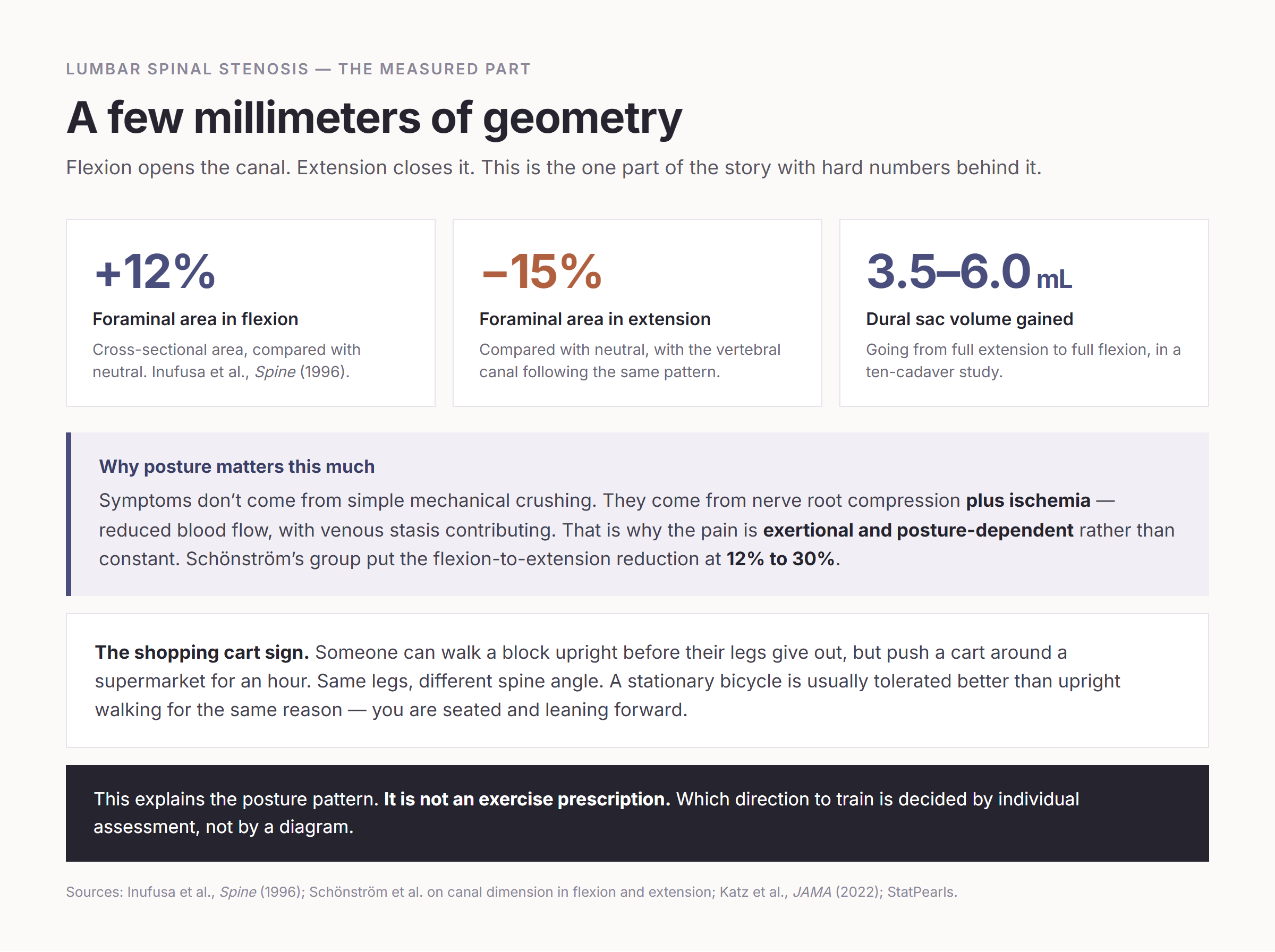
Symptoms don’t come from simple mechanical crushing. They come from nerve root compression plus ischemia — reduced blood flow, with venous stasis contributing. That’s why the pain is exertional and posture-dependent rather than constant. It’s a plumbing problem as much as a pinching problem.
The signature symptom is neurogenic claudication: pain, heaviness, numbness or fatigue in the buttocks and legs that comes on with standing and walking, and eases within minutes of sitting down or bending forward. Numbness in stenosis typically involves the whole leg rather than a single nerve-root stripe. Roughly 43% of patients report weakness.
For scale: an estimated 103 million people worldwide are affected (Katz, JAMA 2022), and symptomatic prevalence climbs steeply with age — in a Japanese population study, 1.9% at 40–49, 4.8% at 50–59, 5.5% at 60–69, and 10.8% at 70–79. In the United States, stenosis is the leading preoperative diagnosis for spine surgery in adults over 65, with roughly 600,000 procedures a year.
Why leaning forward helps — and by how much
This is the one part of the story with hard numbers behind it.
Flexion opens the canal. Extension closes it. Inufusa et al. (Spine, 1996) measured it directly: foraminal cross-sectional area was 12% greater in flexion and 15% smaller in extension compared with neutral, with the vertebral canal following the same pattern. Schönström’s group found the reduction from flexion to extension ranged from 12% to 30%. In a ten-cadaver study, dural sac volume increased by 3.5 to 6.0 mL going from full extension to full flexion.
A few millimeters of geometry, and a leg that works or doesn’t.
The shopping cart sign
That mechanism has a name in the exam room. The “shopping cart sign” describes someone who can walk a block upright before their legs give out, but can push a cart around a supermarket for an hour. Same legs, different spine angle.
The related signs follow the same logic:
- Simian stance — a habitual slightly flexed trunk and bent knees
- Better tolerance of a stationary bicycle than of upright walking, because you’re seated and leaning forward
- Symptoms worst with standing, walking, walking downhill, and lying flat; better sitting or leaning on something
Photo: Charlie Quirk / Pexels
Stenosis vs. a herniated disc — opposite posture signatures
If you bring one observation to your appointment, bring this one. The two conditions respond to posture in opposite directions.
| Lumbar spinal stenosis | Lumbar disc herniation | |
|---|---|---|
| Typical age | Older adults, rising steeply after 50–60 | Often 30s–50s |
| Onset | Gradual, months to years | Often sudden, tied to a movement or lift |
| Worse with | Extension — standing, walking, lying flat | Flexion — sitting, bending forward |
| Better with | Flexion — sitting, leaning on a cart, cycling | Often standing or walking; varies |
| Symptom pattern | Both legs, asymmetric; usually the whole leg | Usually a single nerve-root stripe |
| Cough, sneeze, straining | Usually no worse | Worse — a useful discriminator |
| Straight leg raise | Positive in only about 10% | Frequently positive |
| Walking-induced pain | The hallmark | Less characteristic |
Bending forward is the disc patient’s enemy and the stenosis patient’s friend. That’s a genuinely useful clue.
But it is a clue, not a diagnosis — and here’s the catch that matters later. Older adults very often have both. Degenerative discs and stenosis live in the same spine, which is exactly why blanket exercise rules aimed at a label tend to fall apart on real people.
Neurogenic vs. vascular claudication — please don’t sort this out yourself
Leg pain that comes on with walking has a second common cause in this age group: peripheral artery disease. Confusing the two is genuinely dangerous, in both directions.
| Feature | Neurogenic (stenosis) | Vascular (PAD) |
|---|---|---|
| Where | Above the knees — buttocks, thighs | Below the knees — classically the calf |
| Relieved by | Sitting or bending forward | Standing still, no posture change needed |
| Trigger | Can start from standing alone; distance varies | A consistent, reproducible walking distance |
| Cycling | Usually well tolerated | Provokes symptoms |
| Walking uphill / leaning forward | Easier | Harder |
| Pedal pulses | Normal | Diminished or absent |
| Key test | MRI, after a clinical diagnosis | Ankle-brachial index; ≤ 0.90 signifies PAD |
| Skin and limb | Normal | Hair loss, shiny or cool skin, slow-healing wounds |
One study found that the combination of symptoms triggered by standing plus relief with sitting plus pain above the knee plus a positive shopping cart sign carried a likelihood ratio of 13 for neurogenic claudication. In the JAMA Rational Clinical Examination review (Suri et al., 2010), a wide-based gait also carried LR 13.
Now the reason I’ve framed this as questions for a clinician rather than a self-test.
PAD is common and quietly under-recognized in exactly this age group — prevalence of 8.0–9.0% at ages 65–74 and 16.8% at 75 and older among Medicare beneficiaries. Only about 10% of people with PAD have classic claudication; 30–60% report no exertional leg symptoms at all. And PAD carries a two- to threefold increase in cardiovascular morbidity and mortality, because it is systemic atherosclerosis wearing a leg costume.
Filing a PAD patient under “just stenosis” means missing a treatable cardiovascular diagnosis. The reverse error wastes months. And both conditions can sit in the same person at the same time.
So bring the table. Don’t rule with it.
“My MRI says severe stenosis” is not a diagnosis
This is the correction I’d most like to hand to anyone holding an alarming radiology report.
In the Wakayama Spine Study (Ishimoto et al., Osteoarthritis and Cartilage, 2013), 938 general-population participants — mean age 66.3 — were imaged with mobile MRI. 77.9% had more than moderate central stenosis. 30.4% had severe central stenosis.
And of those with severe central stenosis, only 17.5% actually had symptoms.
The rest of the literature says the same thing from other angles:
- Boden et al.: 21% of asymptomatic people over 60 have lumbar spinal stenosis on MRI.
- Framingham (Kalichman et al., Spine J, 2009): 19–47% of Americans over 60 show anatomic stenosis on imaging, depending on which criteria you use. That range is itself the story — there is no single agreed radiologic definition.
- Brinjikji et al. (AJNR, 2015), reviewing 3,110 asymptomatic people: disc degeneration runs from 37% at age 20 to 96% at age 80. Degenerative findings are normal aging.
- A 2022 review update states it flatly: symptom intensity is “not directly correlated” with the degree of stenosis on MRI.
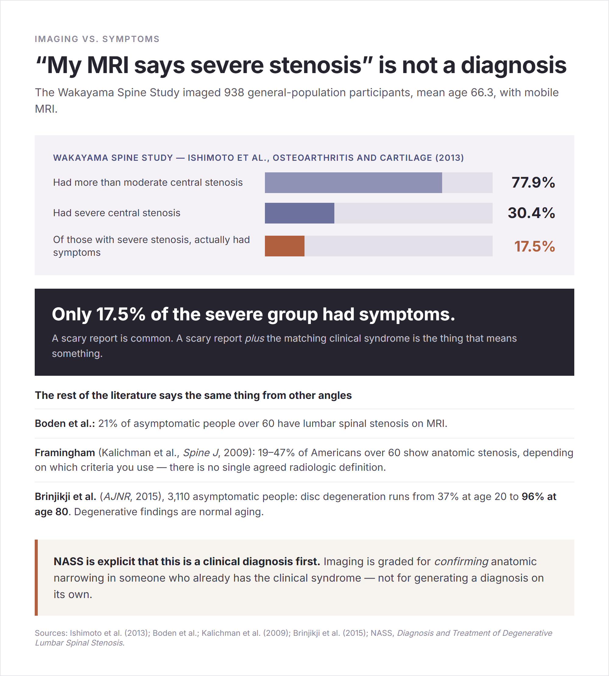
NASS is explicit that this is a clinical diagnosis first. Its history and physical recommendation is to consider stenosis in older patients with buttock or leg symptoms that worsen with walking or standing and improve with sitting or bending forward. Imaging is graded for confirming anatomic narrowing in someone who already has the clinical syndrome — not for generating a diagnosis on its own.
Which means the sentence “my scan shows severe stenosis, so I need surgery” skips the only step that actually decides anything.

Photo: Ivan S / Pexels
Is it going to keep getting worse?
Not necessarily, and this deserves saying because the assumption drives a lot of anxious decisions.
NASS concluded the natural course is favorable in one-third to one-half of people with mild-to-moderate stenosis. Roughly a third improve, most stay stable, and a 10-year longitudinal study found 60% did not progress dramatically. Rapid neurological deterioration is rare, and surgery for stenosis is “almost always elective.”
The other side of the ledger, stated just as plainly: 20–40% of people with mild-to-moderate stenosis do end up having surgery within 10 years.
Both of those are true. Neither is a prediction about you.
What exercise actually helps
Here’s where I have to be careful, because this is the section most articles get wrong by sounding certain.
The strongest evidence is for a package, not a movement. The 2021 clinical practice guideline in The Journal of Pain (Bussières, Cancelliere, Ammendolia et al.), built with GRADE methodology, recommends a multimodal approach: education, advice and lifestyle change, and behavioral techniques combined with home exercise, manual therapy and rehabilitation. The exercise components it names are stretching, strength training, cycling, and body-weight-supported treadmill walking — delivered as an “individually tailored supervised and home exercise program.” It recommends against single-modality approaches as primary treatment.
The 2022 updated systematic review (Ammendolia et al., BMJ Open, 44 randomized trials) sorts the pieces by evidence quality:
| What | Evidence | Finding |
|---|---|---|
| Manual therapy + exercise | Moderate | Clinically important short-term gains vs. medical care or group exercise |
| Manual therapy + education + exercise with a cognitive-behavioral approach | Moderate | Better walking distance, immediate to long term |
| Supervised PT vs. home exercise alone | Low | Supervised better |
| Aquatic exercise | Low / very low, single trial | Better than PT using heat and TENS |
| Stationary cycling | Low | Equivalent to body-weight-supported treadmill walking |
| Oral medications (pregabalin, gabapentin, oxymorphone) | Very low | No meaningful benefit over placebo |
A European Spine Journal review in 2022 added that exercise is significantly better than no exercise, while admitting it was “unable to draw conclusions on which physical therapy treatment is superior.”
And there is one program with real numbers: Ammendolia’s six-week, twice-weekly “boot camp” of manual therapy, education and home exercise. 81% of participants hit the walking improvement threshold at 12 months, and in one trial 85% of moderately disabled patients improved walking distance after six weeks, sustained at a year.
The “never extend” rule, and why it isn’t one
Now the correction I started with.
Directionally, the conventional advice is reasonable. Most people with stenosis feel better in flexion and worse in extension, the anatomy explains why, and a flexion-biased program is a sensible default starting point.
But “extension and McKenzie exercises are contraindicated in stenosis” is a clinical rule of thumb, not a guideline recommendation. Four things, all worth knowing:
- NASS grades physical therapy and exercise as Grade I — insufficient evidence to recommend for or against as a stand-alone treatment. It makes no statement that extension is contraindicated.
- The 2021 guideline contains no prohibition on extension, and its whole posture is “individually tailored.” The 2022 Ammendolia review doesn’t compare flexion-based against extension-based programs at all.
- A survey of UK primary care physiotherapists found most routinely use flexion-based exercise, “reflecting recommendations based on theoretical benefits, but for which trial evidence is lacking.” That sentence is the honest state of the field.
- McKenzie/MDT is not an extension protocol. It is a classification system driven by directional preference. It’s associated with extension because 67–85% of general low back pain patients happen to prefer extension — and the method’s own literature says that when the extension response is unfavorable, a flexion-based regimen should be used instead. There is even a published case report of a 64-year-old man with stenosis who failed a flexion-based program and responded to an extension-based one after directional-preference testing.
So please don’t take this as permission to go do back bends. Take it as the opposite of a rule in either direction.
End-range repeated extension — prone press-ups, standing back bends, sustained backward bending — commonly provokes stenosis symptoms, and holding off until someone assesses you is sensible. Not because a guideline forbids it, but because it frequently hurts and there’s no evidence you’re missing out.
And flexion isn’t risk-free either. A trial in the 2022 review had a flexion-exercise arm that underperformed manual therapy plus unloaded treadmill walking. Coexisting disc pathology can be aggravated by aggressive flexion. And repeated loaded spinal flexion is a recognized concern for vertebral compression fracture in people with osteoporosis — a group that overlaps heavily with this one. (That last point is a general osteoporosis caution rather than something verified in a stenosis-specific guideline, so treat it as a question to raise with your clinician, not a prohibition.)
The actual rule is simple to state and impossible to shortcut:
Train the direction that reduces your symptoms — determined by individual assessment, not by the label on an MRI report.
What’s commonly on the list
These are the approaches that appear in evidence-consistent programs. Frequency, sets and progression are a clinician’s call, and no source supports a specific protocol, so I’m not going to invent one.
- Interval walking — walk to the point before symptoms start, sit for a minute or two, resume. Repeated bouts beat pushing through. A cart, walker or trekking poles extend the distance considerably for many people.
- Stationary cycling — the best-tolerated aerobic option for most, with direct trial support.
- Aquatic exercise or pool walking — buoyancy unloads the spine; positive but very-low-quality evidence.
- Body-weight-supported treadmill walking — supervised, and roughly equivalent to cycling.
- Core, trunk and hip strengthening — named as guideline components. Be honest about it though: the moderate evidence for core strengthening is specifically in post-surgical recovery, and stand-alone core work is not a proven cure.
- Education and pacing — the component with the strongest evidence signal, and the one consumer articles leave out most often.
A useful thing to track is the self-paced walking test — how far you get before symptoms stop you. It’s the standard outcome measure in this population, and it beats “my back feels about the same.”

Photo: Kampus Production / Pexels
Injections and surgery — where the evidence genuinely conflicts
I’m not going to resolve these for you, because the literature hasn’t.
Epidural steroid injections: three positions, all published
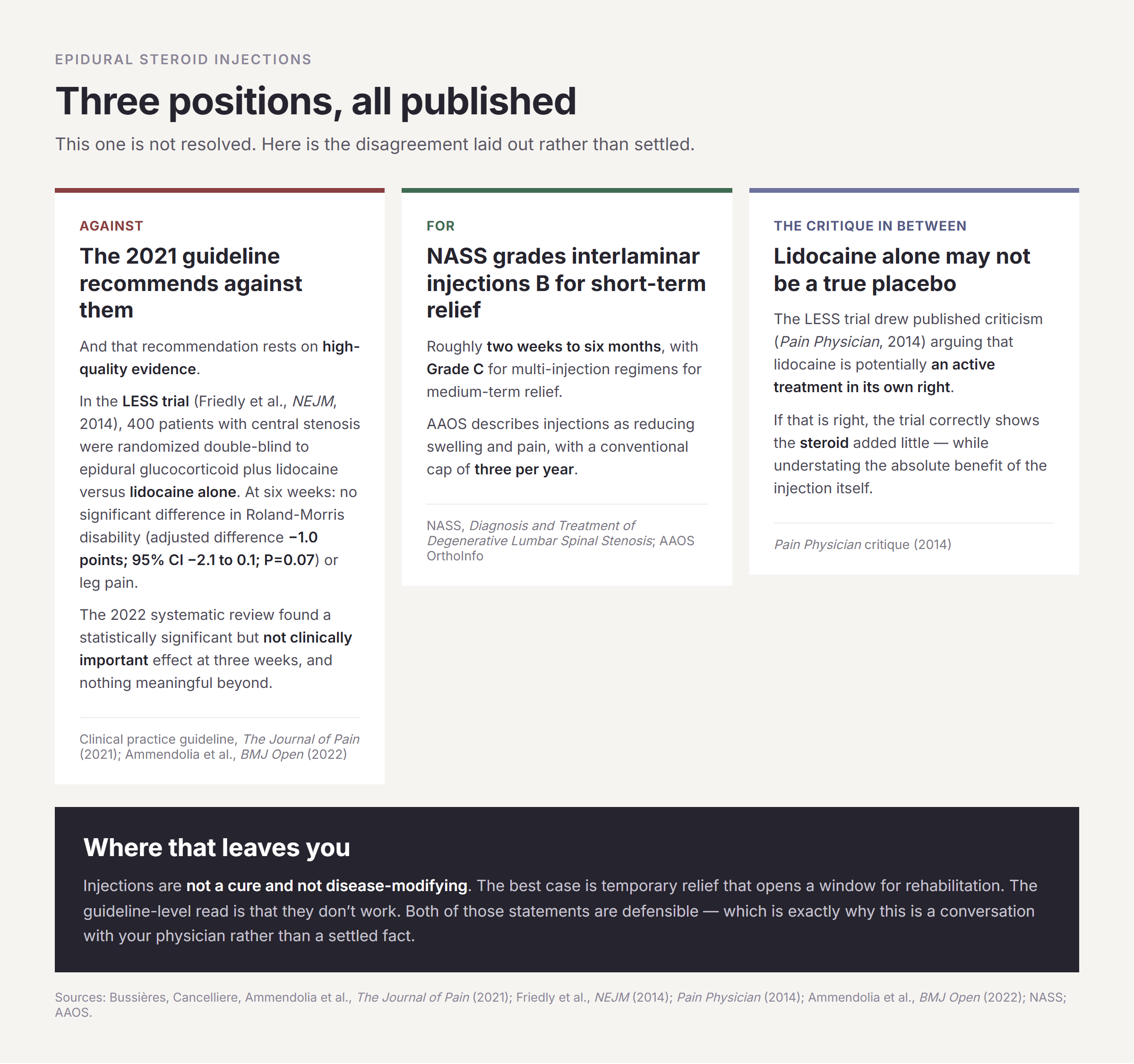
Against. The 2021 clinical practice guideline recommends against epidural steroid injections, and that recommendation rests on high-quality evidence. In the LESS trial (Friedly et al., NEJM, 2014), 400 patients with central stenosis were randomized double-blind to epidural glucocorticoid plus lidocaine versus lidocaine alone. At six weeks there was no significant difference in Roland-Morris disability (adjusted difference −1.0 points; 95% CI −2.1 to 0.1; P=0.07) or leg pain. The 2022 review found a statistically significant but not clinically important effect at three weeks and nothing meaningful beyond.
For. The NASS guideline gives Grade B to interlaminar injections for short-term relief, roughly two weeks to six months, and Grade C to multi-injection regimens for medium-term relief. AAOS describes injections as reducing swelling and pain, with a conventional cap of three per year.
The critique in between. The LESS trial drew published criticism (Pain Physician, 2014) arguing that lidocaine alone isn’t a true placebo — it may be an active treatment in its own right. If that’s right, the trial correctly shows the steroid added little while understating the absolute benefit of the injection.
Where that leaves you: injections are not a cure and are not disease-modifying. The best case is temporary relief that opens a window for rehabilitation. The guideline-level read is that they don’t work. Both of those statements are defensible, which is exactly why this is a conversation with your physician rather than a settled fact.
Surgery: real benefit, real trade-offs, narrowing over time
Surgery is typically considered after three to six months of conservative care for persistent, function-limiting symptoms. NASS suggests decompression for moderate-to-severe symptoms, and decompression alone for leg-dominant symptoms without instability.
The trials, side by side:
- SPORT (Weinstein et al., NEJM, 2008; 4-year results 2010; 10-year review 2019): in intent-to-treat analysis, no difference — driven by 43% crossover from the non-surgical arm. As-treated analyses favored surgery from six weeks through four years, and between years 4 and 8 the gap narrowed as both groups stabilized.
- Delitto et al. (Annals of Internal Medicine, 2015): 169 surgical candidates aged 50+, randomized to surgery or physical therapy. Similar outcomes at two years. Also with heavy crossover — 57% of the PT group went on to surgery.
- Cochrane (Zaina et al., 2016): five trials, 643 participants. “No clear benefits were observed with surgery versus non-surgical treatment.” And the number that should be in every consent conversation — adverse events in 10–24% of surgical participants, including fracture, stroke and death, against no side effects reported for any conservative treatment.
- Fusion: randomized trials by Försth (2016) and Ghogawala (2016) found no significant difference between decompression alone and decompression plus fusion for most stenosis patients, while fusion added blood loss, operative time and hospital stay. For isolated stenosis without spondylolisthesis or scoliosis, fusion has not shown added benefit.
The optimistic figures exist too and deserve equal air: liberal laminectomy is described as benefiting close to 80% of patients with severe stenosis, and AAOS reports good-to-excellent results in the majority, with desk work resumed in weeks.
So the accurate summary isn’t “surgery wins” and it isn’t “never operate.” It’s this: surgery tends to produce faster relief in the first one to four years for well-selected patients with moderate-to-severe symptoms who have failed conservative care, that advantage narrows by six to eight years, one high-quality trial found physical therapy equivalent at two years, and the safety ledger clearly favors conservative care.
The editorial accompanying the Delitto trial said it best in its title: “Informed Patient Preferences Should Weigh Heavily.”

Photo: Funkcinės Terapijos Centras / Pexels
Stop reading and go — the cauda equina emergency
Everything above assumes you have ordinary stenosis. There is one pattern that is not ordinary.
If you develop new trouble controlling your bladder or bowels, new numbness in the groin or saddle area, or sudden weakness in both legs, go to the emergency department the same day. Do not wait for a clinic appointment, do not “sleep on it,” and do not try exercises. Call 911 or have someone drive you.
That cluster can indicate cauda equina syndrome — compression of the bundle of nerve roots below the end of the spinal cord, and a neurosurgical emergency. New bladder dysfunction appears in up to 92% of cases and saddle anesthesia in up to 93%. Back pain and sciatica accompany it in as many as 97% of cases, but pain alone is not the red flag — the bladder, bowel, saddle and bilateral-weakness cluster is.
Timing is the whole reason for the urgency. Better prognosis is associated with decompression within 48 hours of presentation, and MRI should ideally happen within one hour of arrival. Deficits can become permanent.
It is genuinely rare — roughly 1 in 30,000 to 1 in 100,000 people per year — which is precisely why the specific symptom list is more useful than generalized fear.
A second, non-emergency tier still deserves a prompt appointment: progressive foot drop, worsening balance or falls, unexplained weight loss, fever with back pain, a history of cancer, or pain that is severe at rest and at night.
What I’d actually do with all this
If I were sitting with the report and the leg pain, here’s the order I’d take it in.
The posture pattern is the most informative thing I own — better in flexion, worse standing upright, better on a cart or a bike. That’s worth describing precisely to a clinician, along with how far I can walk before it starts.
The MRI is context, not a verdict. 77.9% of a general population over 60 had more than moderate stenosis, and only 17.5% of the severe group had symptoms. A scary report is common. A scary report plus the matching clinical syndrome is the thing that means something.
Leg pain with walking gets a circulation check, not an assumption. Pulses, and an ankle-brachial index if there’s any question.
And the exercise question doesn’t have a universal answer, which was the whole surprise for me. Most people do better with a flexion bias. Some don’t. The evidence supports the package — education, tailored exercise, manual therapy, pacing — far more than it supports any particular movement. That means an actual assessment, by a physical therapist or physician, rather than a rule copied off a diagram.
I came in looking for the exercise you’re not allowed to do. What I found instead was a field that has measured the anatomy to the millimeter and is still, in 2026, mostly guessing about the movements — and that’s worth knowing before you let anyone sound too certain about your spine.
Medical disclaimer. This article is general health information, not medical advice, and it does not establish a patient–clinician relationship. Lumbar spinal stenosis is diagnosed clinically by a physician; imaging findings alone do not make the diagnosis and do not determine treatment. Do not start, stop, or change any medication based on this article — medication choice, including anything mentioned by class here, is a prescriber’s decision.
Exercise disclaimer. Exercise for spinal stenosis should be individually assessed and prescribed. An approach that helps one person can worsen another, and no article can tell you which direction your symptoms respond to. Get assessed by a physical therapist or physician before starting a program, and stop and get reassessed if your symptoms worsen, spread further down the leg, or if new weakness or numbness appears. If you have osteoporosis or low bone density, raise that specifically before starting loaded flexion work.
Emergency. New trouble controlling your bladder or bowels, new numbness in the groin or saddle area, or sudden weakness in both legs — go to the emergency department the same day. Do not wait for a clinic appointment, do not “sleep on it,” and do not try exercises. Call 911 or have someone drive you.
References
- NASS — Diagnosis and Treatment of Degenerative Lumbar Spinal Stenosis (2011 update)
- Bussières, Cancelliere, Ammendolia et al. — Non-Surgical Interventions for Lumbar Spinal Stenosis Leading To Neurogenic Claudication: A Clinical Practice Guideline, The Journal of Pain (2021)
- Ammendolia et al. — updated systematic review of non-operative treatment, BMJ Open (2022); European Spine Journal physiotherapy review (2022)
- Zaina et al. — Surgical versus non-surgical treatment for lumbar spinal stenosis, Cochrane (2016)
- Friedly et al. — LESS trial, NEJM (2014); Pain Physician critique (2014)
- Delitto et al. — Annals of Internal Medicine (2015), with accompanying editorial; Weinstein et al. — SPORT, NEJM (2008), 4-year (2010) and 10-year review (2019)
- Försth (2016) and Ghogawala (2016) — decompression with vs. without fusion
- Ishimoto et al. — Wakayama Spine Study, Osteoarthritis and Cartilage (2013); Kalichman et al. — Framingham, Spine J (2009); Brinjikji et al., AJNR (2015)
- Inufusa et al. — Spine (1996); Schönström et al. — canal dimension in flexion and extension
- Katz et al. — Diagnosis and Management of Lumbar Spinal Stenosis: A Review, JAMA (2022); Lurie & Tomkins-Lane, BMJ
- Suri et al. — JAMA Rational Clinical Examination (2010); Canadian Journal of Surgery (2013) on neurogenic vs. vascular claudication
- StatPearls — Lumbar Spinal Stenosis; Cauda Equina and Conus Medullaris Syndromes; McKenzie Back Exercises; AAOS OrthoInfo — Lumbar Spinal Stenosis
- JAHA (ARIC) and Circulation Research — peripheral artery disease prevalence and symptom patterns